|
AFM
Gallery
|
|
Polymeric materials
|
|
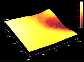
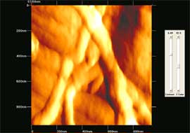
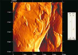
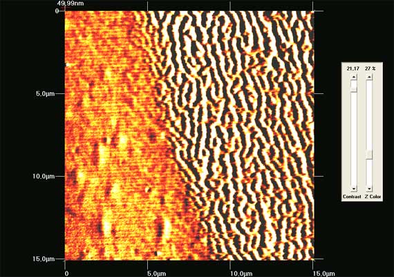
Image between exposed and
non exposed areas of fluoropolymer at 157nm. Materials
were provided by IMEL Demokritos.(zoom)
|

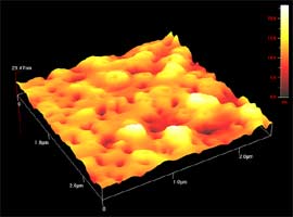
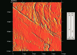
Surface morphology of exposed
area of fluoropolymer at 157nm.
Materials were provided
by IMEL Demokritos.
(zoom)
|
|
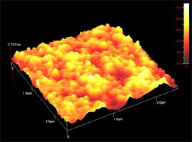
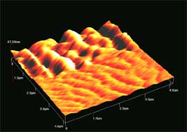
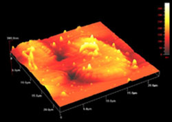
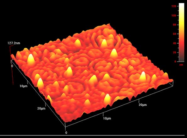
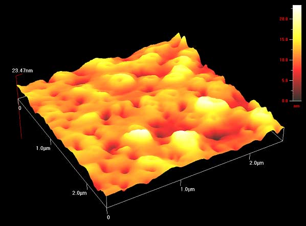
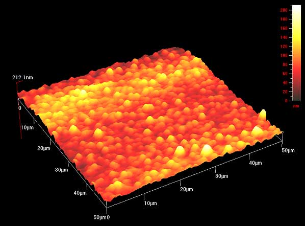
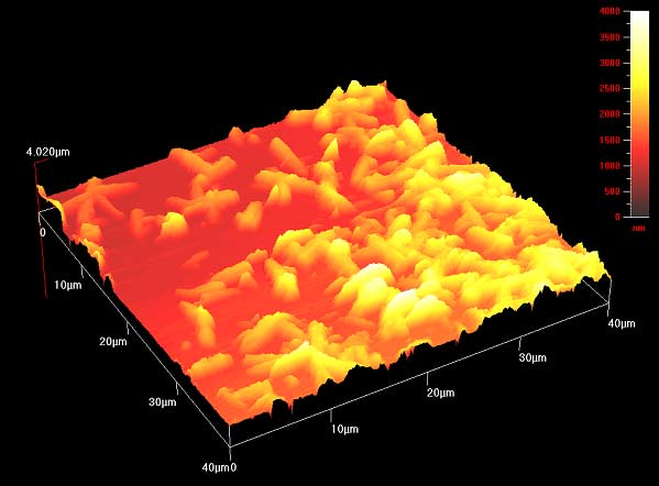
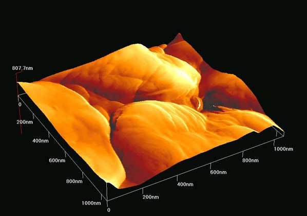
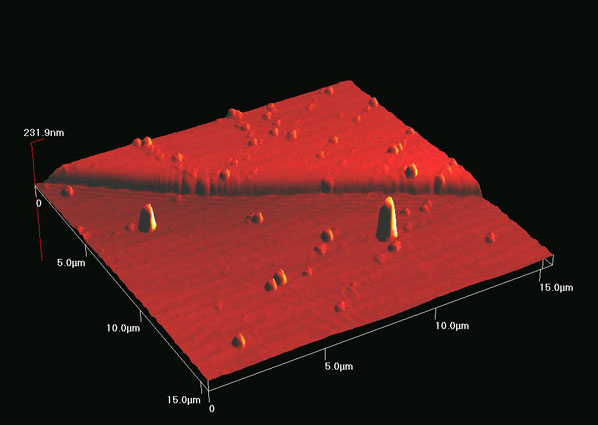
3D image of surface morphology
of fluoropolymer.
Materials were provided by IMEL Demokritos.
(zoom)
|

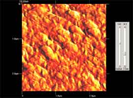
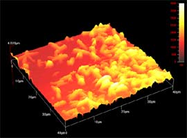
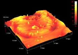
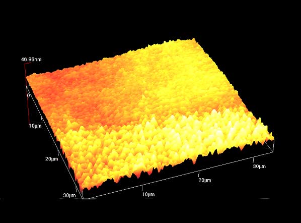
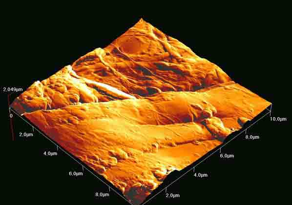
3D surface image of the exposed
area of silicon based polymer at 157nm. Materials
were provided by IMEL Demokritos. (zoom)
|
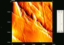
Image between exposed and non exposed areas of silicon based
polymer at 157nm.
Materials were provided by IMEL Demokritos.(zoom)
|
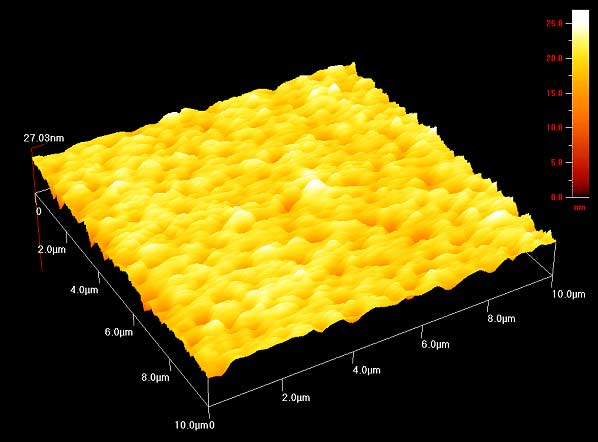
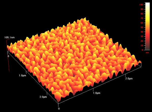
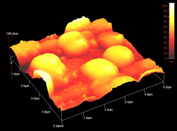
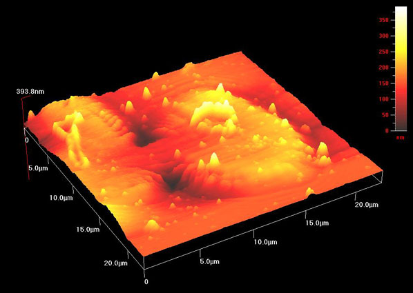
3D image of surface morphology of silicon based co-polymer.
Materials were provided by IMEL Demokritos. (zoom) |
3D image of surface morphology of silicon
based co-polymer. Materials were
provided by IMEL Demokritos.
(zoom)
|

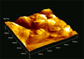
3D image of surface
morphology of silicon based co-polymer.
Materials were provided
by IMEL Demokritos.
(zoom)
|
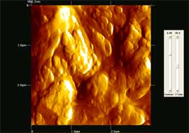
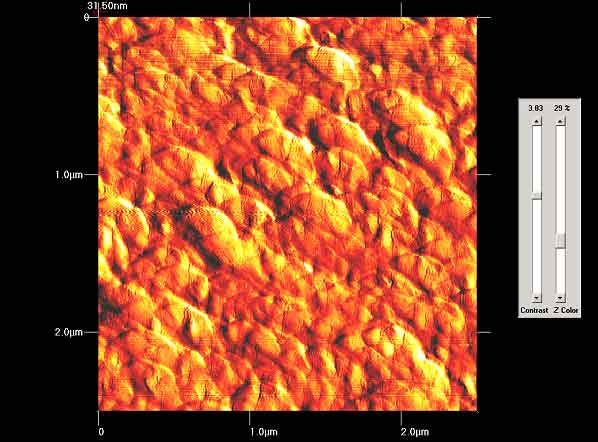
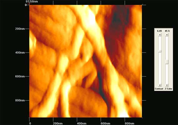
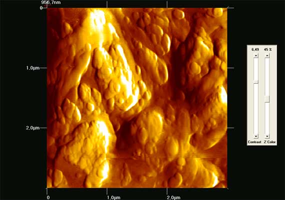
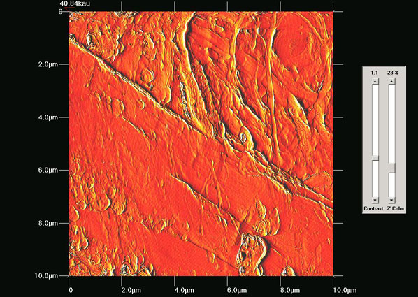
2D image of surface
morphology of silicon based co-polymer. Materials
were provided by IMEL Demokritos.
(zoom)
|
3D image between exposed and
non exposed areas of Teflon film at 157nm. (zoom)
|
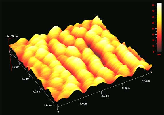
3D image of surface morphology of Teflon
film .
(zoom)
|

2D image between exposed and
non exposed areas of Teflon film at 157nm.(zoom)
|
|
|
3D image of Si wafer coated with Ta layer
using laser at 157nm.(zoom)
|

High resolution 3D image of
Si wafer coated with Ta layer using laser at 157nm. (zoom)
|
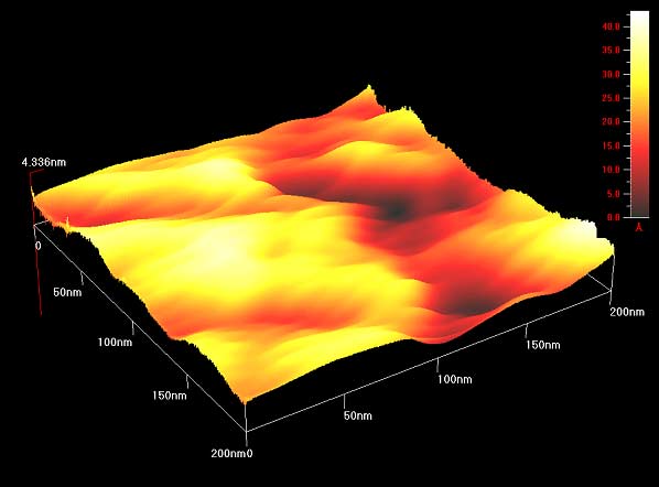
3D image of surface roughness of CaF2
window.(zoom)
|

Quality evaluation of Au sputtered
films.(zoom)
|
|
|
3D image of DNA crystals (Adenine).(zoom)
|
2D image of DNA crystals (Adenine).(zoom)
|
2D image of DNA crystals (Cytocine).(zoom)
|

3D image of DNA crystals (cytosine). The edge between
the non-irradiated and radiated at 157nm areas is seen. (zoom)
|
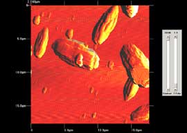
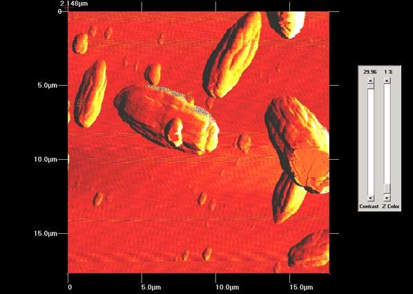
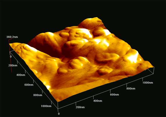
3D image of Ulocladium spore exposed at
157nm.(zoom)
|
3D image of Ulocladium spore
exposed at 157nm.(zoom)
|
|
Historic
Paper
|
2D image of non infected area of historic
paper . (zoom)
|
2D image of non infected
area of historic paper. (zoom)
|

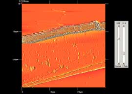
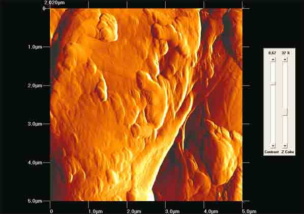
3D image of historic paper area. Infected
by foxing area can be seen on the viewers side of the image. (zoom)
|

3D image of historic paper
areas infected by foxing.
(zoom)
|
2D image of historic paper area infected by foxing. (zoom)
|
2D phase mode image of historic
paper. (zoom)
|
2D image of paper area. (zoom)
|
3D image of paper area.
(zoom)
|
|
|
48,
Vassileos Constantinou Aven. 11635 Athens, Greece
Tel: +30 210 7273840, Fax: +30 210 7273842, email :ccefalas@eie.gr


