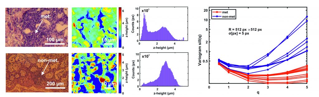
Early ascertainment of metastatic tumour phases is crucial to improve cancer survival, formulate an accurate prognostic report of disease advancement and, most important, quantify the metastatic progression and malignancy state of primary cancer cells with a universal numerical indexing system. This work proposes an early improvement of cancer detection with 97 nm spatial resolution by indexing the metastatic cancer phases from the analysis of atomic force microscopy images of human colorectal cancer histological sections. The procedure applies variograms of residuals of Gaussian filtering and theta statistics of colorectal cancer tissue image settings. The methodology elucidates the early metastatic progression at the nanoscale level by setting metastatic indexes and critical thresholds from relatively large histological sections and categorising the malignancy state of a few suspicious cells not identified with optical image analysis. In addition, we sought to detect early tiny morphological differentiations indicating potential cell transition from epithelial cell phenotypes of low to high metastatic potential. The metastatic differentiation, also identified by higher moments of variograms, sets different hierarchical levels for the metastatic progression dynamic, potentially impacting therapeutic cancer protocols.
Key publications
Nanoscale prognosis of colorectal cancer metastasis from AFM image processing of histological sections.
V. Gavriil, A. Ferraro, A.C. Cefalas, Z. Kollia, F. Pepe, U. Malapelle, C. De Luca, G. Troncone and E. Sarantopoulou,
bioRxiv 2022.05.06.490873
DOI:10.1101/2022.05.06.490873